
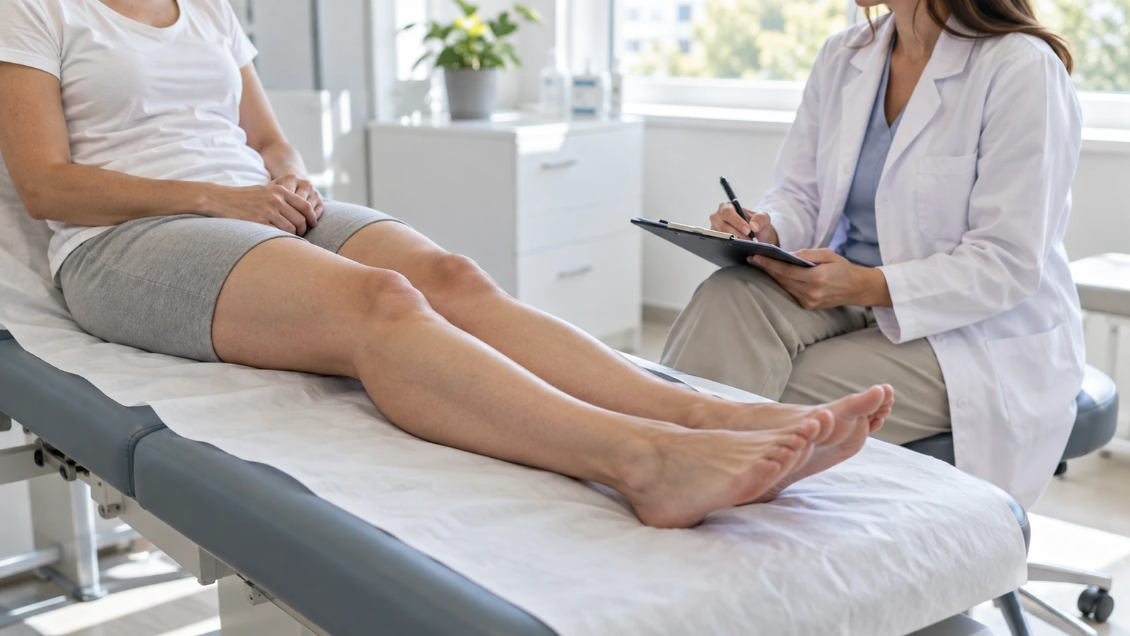
Fat calves rarely have a single explanation. The volume in that area can come from muscle, from fat (because of body build or weight), from fluid held back by the veins or the lymphatic system, or from lipedema affecting the lower leg. Telling them apart matters, because each cause responds differently to exercise and to weight.
You will not find tips here on how to “slim your calves fast”. You will find what we look at in consultation: seven checks you can do at home, a triage table and, for each cause, what genuinely changes and what does not. If you want the whole-leg picture first, it is on our page on the symptoms of lipedema; this article goes one step down, from the whole leg to the calf.
The urgent part first: when a swollen calf cannot wait
Thick calves you have always had are not an emergency. A calf that changes suddenly is. Seek medical attention today if any of these apply to you:
One calf only, swollen, painful, warm or red: this is the typical way a deep vein thrombosis presents, and it almost always affects one side. Do not wait to see whether it goes down.
Leg swelling with breathlessness or chest pain: this can be a clot that has reached the lung. It is an emergency: call your local emergency number (112 in Spain and the EU, 999 in the UK, 911 in the US).
Sudden pain in the calf during exertion, followed by swelling and a limp: this points to a torn calf muscle (gastrocnemius rupture).
A lump behind the knee that suddenly hurts and makes the calf swell: this may be a ruptured Baker’s cyst, but its symptoms are very similar to those of a clot and only an examination can tell them apart.
Red, hot, tight skin that is spreading, with fever or chills: this points to cellulitis, a skin infection that needs antibiotics.
And one that is not an emergency, but not one to let slide either: if, as an adult, one leg alone starts to get bigger for no clear reason, ask for a medical assessment within the next few days. Swelling on one side calls for a search for something that is obstructing the drainage of that leg.
What is inside a calf, and why it bulks out
The calf is the back of the lower leg, between the knee and the ankle. Its outline comes mainly from two muscles, the gastrocnemius and the soleus, which together form the triceps surae and end in the Achilles tendon. Over them lies the subcutaneous fat and, between the tissues, run the veins and lymphatic vessels that carry fluid back up the leg.
Put another way: a calf can be thick because of what lies underneath (muscle), what covers it (fat) or what builds up between the tissues (fluid). Lipedema is a particular case of the second: fat that is out of proportion, symmetrical and painful.
Seven checks you can do at home
None of them gives a diagnosis, but together they point you in a clear direction. Do them on both legs and, if you can, once in the morning and once in the evening: whatever changes over the day is usually fluid.
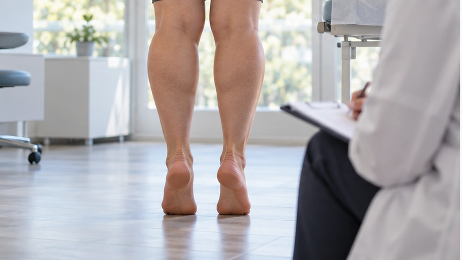
1. Stand on tiptoe. The calf muscle contracts, firms up and stands out. If almost all the volume tightens with you, much of it is muscle. If a soft layer stays on top and does not change, there is fat.
2. Pinch the skin. Take a fold on the inner side of the calf between your thumb and index finger and compare it with one on your forearm. A thick, soft fold is fat; a thin fold over something firm is muscle.
3. Check for pitting. Press with your thumb for a few seconds on the inner shin, where the bone sits just under the skin, then let go. If a dent stays and takes time to fill in, there is fluid.
4. Look at the ankle. If the volume stops abruptly above the ankle, like a cuff, and the foot stays slim, that points toward lipedema. If the foot is swollen too, it points away from it.
5. Pinch the top of the second toe. If you can lift the skin, the Stemmer sign is negative. If it is thickened and will not lift, it is positive and points to a lymphatic component.
6. Squeeze the calf with your hand. In lipedema the fat is tender to pressure, and this is a core criterion: if it does not hurt, it is not called lipedema. A large calf muscle, or fat that simply reflects body build, does not usually hurt when squeezed.
7. Compare one side with the other. Lipedema is symmetrical. If one calf is clearly bigger than the other, the difference has another explanation, even if there is underlying lipedema as well.
A note on the feeling of swelling. Many patients with lipedema feel that their legs “swell” in the evening. In a study comparing women with lipedema and healthy women, that swelling could not be measured objectively, and the authors interpret it as part of the pain experience. If the swelling can actually be seen and leaves a dent, it needs a cause of its own.
Triage table: what points to each cause
What discriminates most is the combination of tenderness to pressure, pitting and the foot.
| Cause | Both or one? | Does it pit? | Does it hurt when squeezed? | Ankle and foot | Weight and exercise |
Muscle (large calf muscle) | Both | No | No | Normal outline | Strength training can make it bigger |
Fat from body build or excess weight | Both, in proportion with the rest of the body | No | No | No marked step | Losing weight reduces the volume, but not “only in the calf” |
Fluid retention or venous insufficiency | Both, or one more than the other | Yes | Heaviness more than pain | Swollen ankle, worse when standing | Improves with the legs raised; the cause needs treating |
Lymphedema | Usually asymmetrical | At first, yes; it may stop pitting | Usually not | Foot and toes involved, Stemmer often positive | Needs assessment and specific treatment |
Lipedema affecting the calf | Always both, symmetrical | Little or not at all | Yes, core criterion | Step above the ankle, foot spared | Weight affects the volume; the disproportion does not go away with diet |
No row is a diagnosis, and several can coexist, for example lipedema with excess weight. The table is there to help you know what to ask, and whom.
When it is muscle: a large calf muscle
If almost all the volume firms up when you stand on tiptoe, the calf does not hurt when squeezed and the ankle has a normal outline, the most likely explanation is muscle. That is anatomy, not disease, and it is not lipedema.
What is worth knowing, honestly, is that calf strength training works the muscle, so it can make the calf bigger, not smaller.
Why are my calves so big?
Often the answer is less clinical than you fear: body build and muscle. Some legs have a large calf muscle by nature, and some store fat in the lower leg, just as others store it on the hips. But if your calves also hurt when you squeeze them, you notice a step above the ankle, or they swell and leave a dent, “it’s genetic” is no longer enough of an explanation, and the checks above will help you sort it out.
Can you slim your calves with exercise?
Not in a targeted way. In one trial, participants trained a single leg for 12 weeks, with hundreds of repetitions per session. They lost fat across the body as a whole, mainly in the arms and trunk, but not in the trained leg. Exercise helps you lose fat overall and, in lipedema, helps reduce pain; what it does not do is choose where the fat comes off.
When it is fat: body build and weight
Fat that reflects body build, or the fat that comes with excess weight, is distributed proportionally: it also increases on the abdomen, back or arms, and the calf follows the trend of the rest. It does not hurt when pressed. The reference clinical guideline puts it this way: the obesity that can occur in any woman is proportional and also affects the trunk, unlike lipedema.
There is an in-between case worth naming: a symmetrical disproportion of fat in the legs that does not hurt. The guideline calls it lipohypertrophy and is explicit: without symptoms, lipedema should not be diagnosed (Recommendation 2.2, 100% consensus).
What changes and what does not: when weight goes up, leg volume always goes up; when weight comes down in overweight or obesity, leg volume usually comes down too. What does not exist is weight loss aimed at the calf.
How do you lose calf fat?
It depends on what is making your calves thick, which is why the first honest answer is to find out. If it is fat from weight, losing weight with an appropriate diet and regular movement also reduces the volume of the legs. If it is fluid, its cause needs treating. If it is muscle, there is nothing abnormal to lose. And if it is lipedema, weight counts, but the disproportion does not go away with diet: at that point the conversation is a medical one.
When it is fluid: retention, veins and lymph
Fluid is recognizable by three features: it leaves a dent, it changes over the course of the day and it improves with the legs raised. The guideline notes that swelling from standing for long periods can occur in any woman and is independent of lipedema.
Venous insufficiency
When the valves in the leg veins do not close properly, blood pools while you stand. It causes heaviness, cramps and pain that gets worse when standing and better with the legs raised, with swelling around the ankle, varicose veins and, over the years, skin changes. If the skin of the lower leg has hardened, we explain it in when the leg swells and turns hard. And if the swelling is concentrated at the ankle, the general causes and the medicines that produce it are covered in why ankles swell.
Lymphedema
Lymphedema is protein-rich fluid that the lymphatic system cannot drain. At first it leaves a dent and eases with the leg raised; later the tissue becomes fibrotic and may stop pitting. Unlike lipedema, it affects the foot and toes, the Stemmer sign is usually positive and it is often asymmetrical. Obesity can also bring an associated lymphedema, mainly in the legs. The point-by-point differences are in how to tell lipedema and lymphedema apart.
When it is lipedema: how it shows in the calf
Lipedema (spelled lipoedema in British English) is a disorder of fat distribution that occurs almost exclusively in women and only in the limbs. In the calf it follows a recognizable pattern:
Always in both legs, symmetrically: the guideline describes it as affecting both hips, both thighs or both calves.
The foot is spared. The volume ends above the ankle with a step change in size toward the unaffected area, known as the cuff sign.
It hurts with pressure and touch, with a feeling of heaviness. That is the condition for calling it lipedema.
Disproportion relative to the trunk: the upper body may be slim.
Often, painful fat pads just above or below the knee.
And what it does not have. In pure lipedema, imaging studies have not found edema in the tissue, which is why it pits little or not at all, and the Stemmer sign is negative; it can turn positive if lymphedema is added. Nor is fat that feels “lumpy” a criterion: the guideline advises against using nodular tissue for diagnosis because it lacks validity (Recommendation 2.5, 100% consensus).
Can lipedema affect only the calves?
Yes. The S2k guideline states that the increase in fat can be spread across the thigh and lower leg or affect only the upper or only the lower part, always sparing the feet. That isolated calf pattern is what we call type V on our website. It is also common for the calf to be involved as part of a lipedema that runs down from the hips: in The Advanced Care Study, a survey by our team answered by 1,069 people in Spain (969 included in the analysis), the most commonly reported type was the one running from the hips to the calves (type III in the classification the study used), and the calves were the area most often operated on among the patients who had had surgery.
Can one calf bigger than the other be lipedema?
Not as the explanation for the difference. Lipedema is symmetrical, so a clear asymmetry calls for another cause: the after-effects of an old thrombosis, a venous problem in that leg, lymphedema or a Baker’s cyst, among others. There may be underlying lipedema with another problem on top that explains why one side is bigger. If the asymmetry is recent or painful, go back to the warning signs at the start.
Type V: what it means and what it does not
Type classifications describe where the fat is, not how far the disease has advanced. Just as the stage describes a state of the tissue, not a verdict, the type is a map, not a severity scale.
It pays to be precise about the number, because it is not universal. The S2k guideline brings together four type systems from different authors in a single table. Lipedema “only in the calf” as type V appears in the system by Kruppa and colleagues (2020); in the one by Beltran and Herbst (2017), type V means something else, lipedema of the arms and legs, mainly the legs. Because of that disparity, the guideline does not endorse any of them and asks for morphology to be used descriptively and never as a measure of severity (Recommendation 2.3, 100% consensus).
We use the numbering I to V as a practical way of naming where the fat sits, and we explain it with our own illustrations in stages and types of lipedema. For your calves, what matters is not the number but whether the examination confirms or rules out lipedema.
If it is lipedema, what changes with weight and exercise
Two half-truths circulate here. The first is that “lipedema fat does not respond to diet”. The guideline qualifies this: where there is overweight or obesity, losing weight with an appropriate diet also reduces leg volume (Recommendation 10.2, 100% consensus). What diet does not do is erase the disproportion, because the disease arises independently of weight.
The second is that lipedema always gets worse. Not so: the guideline asks for it not to be understood as a disease that advances as a matter of principle (Recommendation 2.10, 94.4% consensus). With a stable weight it can remain stable for years; what can make it worse are individual factors, such as weight gain or hormonal changes. And pain does not go hand in hand with volume: a very thick calf may hurt little, and a modest one a great deal.
Exercise works in a similar way. Moving, ideally while wearing compression, is a central part of reducing pain (Recommendation 7.2, 100% consensus). What you should not expect is a slimmer calf: in the studies the guideline reviews, training consistently reduced pain, but its effect on leg volume was mixed (no significant change in one study, a reduction in another). Which activity suits each case is covered in exercises for lipedema.
What an assessment involves
If you have spent years hearing that your calves “are just like that” or that “it’s fluid retention”, and it does not add up, what you need is not a treatment: it is a diagnosis. Lipedema is diagnosed clinically (Recommendation 2.12, 100% consensus), through your history and an examination. At our clinic in Valencia the assessment includes pitting and the Stemmer sign on both legs, tenderness to pressure, and Doppler ultrasound to rule out venous disease. If it is not lipedema, we will tell you: ruling things out is part of the job. The team sees international patients in English, and the practicalities of traveling are set out on our page for international patients.
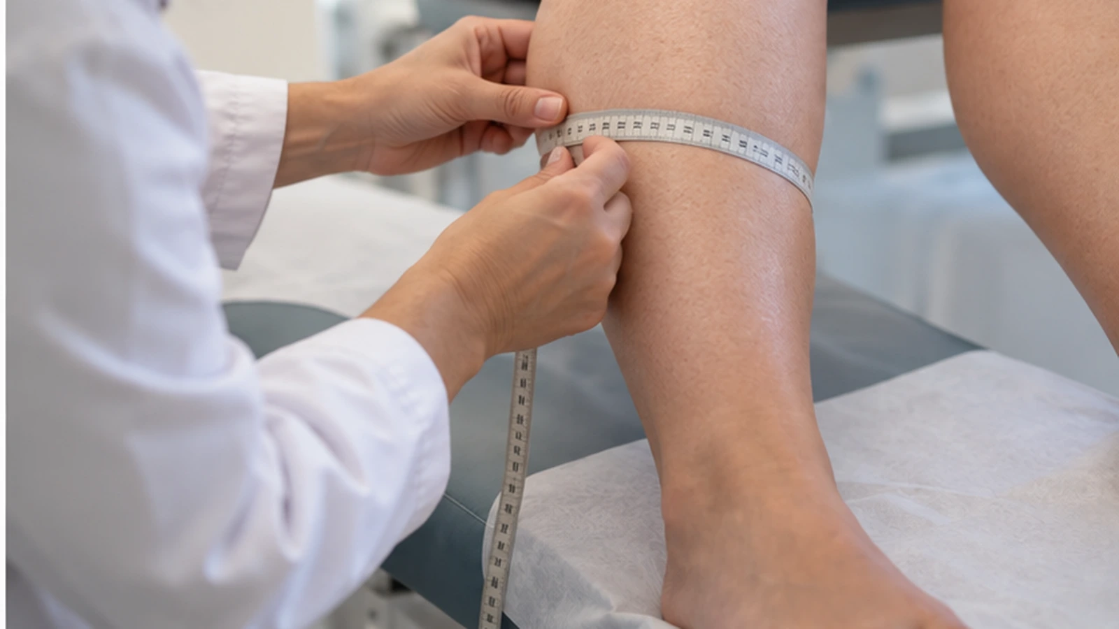
The step-by-step process, and which tests make sense, is explained on our lipedema diagnosis page.
Sources and references
Guideline S2k Lipoedema, AWMF 037-012, version 5.0 (2024). For the definition, symmetry and sparing of the foot, the step change in size, pain as a criterion, lipohypertrophy, proportional obesity, orthostatic swelling, the Kaposi-Stemmer sign, the absence of edema in pure lipedema, the table of types (Table 1) and Recommendations 2.2, 2.3, 2.5, 2.10, 2.12, 7.2 and 10.2.
Peripheral Lymphedema: 2020 Consensus Document, International Society of Lymphology. For the stages of lymphedema, the loss of pitting and the workup of swelling in a single limb.
Anatomy, Bony Pelvis and Lower Limb: Gastrocnemius Muscle. StatPearls.
Ramírez-Campillo R, et al. Regional fat changes induced by localized muscle endurance resistance training. J Strength Cond Res, 2013;27(8):2219-24. PubMed.
Carballeira Braña A, Poveda Castillo J. The Advanced Care Study: Current Status of Lipedema in Spain. Int J Environ Res Public Health, 2023;20(17):6647. PubMed.
Content prepared by the medical team at Lipedema Advanced Care based on the S2k lipedema guideline (AWMF 037-012, 2024 version) and the sources cited.
Published in October 2026.
This page is for information purposes and does not replace a medical consultation. Lipedema is diagnosed clinically, and the diagnosis must be made by an experienced professional. Call your local emergency number now (112 in Spain and the EU, 999 in the UK, 911 in the US) if leg swelling comes with breathlessness or chest pain. Seek medical attention the same day if you have one calf only that is swollen, painful, warm or red; sudden pain in the calf during exertion, with swelling; a lump behind the knee that hurts and makes the calf swell; or red, hot, tight skin with a fever. If one leg alone starts to get bigger for no clear reason, ask for an assessment within the next few days.

