
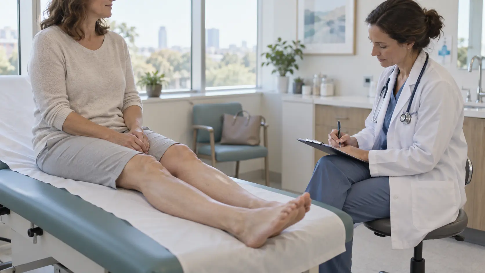
A leg that swells is one thing. A leg that is hard is another. If you press and no dent stays, or if the skin around your ankle has turned stiff and dark, what is underneath is no longer water: the tissue has changed.
We have ordered this around the two questions that actually discriminate: one leg or both? and does it still pit? And we will say the conclusion in the first line, because it is the opposite of what you will read elsewhere: one hard swollen leg is very rarely lipedema.
The urgent part first: when this cannot wait
A single leg, swollen and tense, is how several conditions present that are dealt with the same day. If you recognise any of these, seek medical attention today:
One leg, suddenly, with calf pain, warmth or redness: this has to rule out a deep vein thrombosis. Up to half of acute cases may show no specific signs, so the threshold for getting seen has to be low.
Swelling with breathlessness or chest pain: this can be a pulmonary embolism. It is an emergency.
Red, hot, painful skin with fever or chills: this points to erysipelas or infectious cellulitis. In erysipelas the red edge is well demarcated and raised. It needs an antibiotic, not a cream.
Pain out of proportion, with the leg tense and hard as wood, after a knock or intense exertion: this is acute compartment syndrome. Pain out of proportion to the injury and a palpably tense compartment are what define it. It is a surgical emergency and the window is measured in hours, and it is the one presentation where a hard swollen calf on its own is the alarm.
A wound that will not close over the hardened area.
The two questions that order everything else
Before naming any disease, two things get answered: laterality (one leg or both) and consistency (does it pit or not). Combined they give four scenarios, and each sends you somewhere different.
| Scenario | What gets ruled out first |
One leg, soft (it pits) | Deep vein thrombosis if sudden. If longstanding: post-thrombotic syndrome or venous obstruction. |
One leg, hard (no longer pits) | Fibrotic lymphoedema, the sequel of an old thrombosis, or advanced venous insufficiency. Lipedema is out of the picture. |
Both legs, soft (they pit) | General causes: heart, kidney, liver, thyroid, medication, heat, standing. |
Both legs, hard and tender to pressure | This is where lipedema comes in, and the mixed picture with a lymphatic component. |
Look at the second row. One hard leg points away from lipedema: the reference guideline describes it as an always symmetrical disorder of fat distribution.
The pitting sign: the examination that changes everything
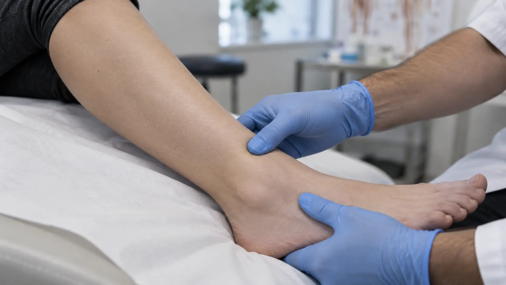
Pitting is the dent left behind when you press and lift your finger. How to check it: place your thumb where the bone sits just under the skin, such as the inner shin, press firmly and hold for several seconds. Lift your finger and look.
If a dent stays and takes time to fill in: there is free fluid in the tissue. The cause lies in the veins, in fluid balance, or in a lymphatic system not yet fibrosed.
If no dent stays and the tissue feels dense, rubbery or woody: what is underneath is no longer water, but fatty or fibrosed tissue. This is what changes the treatment.
How long do you have to press to see whether a leg pits?
Several seconds of sustained pressure over bone, not a quick tap: with a quick tap almost no oedema leaves a mark. Do it on both legs at the same point: if one pits and the other does not, that in itself is a finding.
One hard swollen leg: what to rule out before anything else
Asymmetry carries clinical weight: a general cause (heart, kidney, thyroid) affects both legs at once. When only one swells, the search is for a mechanical obstacle in that leg's drainage.
Thrombosis, post-thrombotic syndrome and venous compression
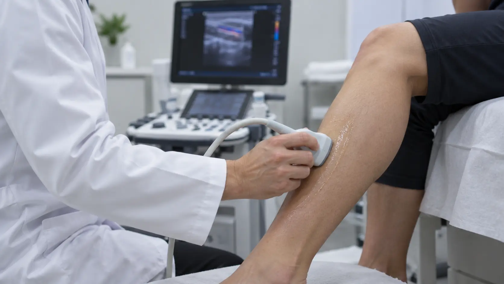
A clot in a deep vein obstructs the return from that leg. In the acute phase it is the emergency from the first section. Months or years later the post-thrombotic syndrome can remain: the vein stays damaged, pressure stays high, and the leg swells, pigments and hardens, typically with a hard swollen calf on that side only. A pelvic vein can also be compressed by a neighbouring structure and impede return on one side. The lipedema guideline says what Doppler ultrasound is for here: identifying alternative vascular diagnoses such as varicose veins, post-thrombotic syndrome or compression syndromes. It does not confirm lipedema: it rules out what imitates it.
Can lipedema affect only one leg?
No. It is symmetrical by definition: it arises only in the limbs, affects both hips, both thighs or both calves, and spares the feet and hands. If one leg is clearly more swollen and harder than the other, another explanation has to be found for that difference, which can coexist with underlying lipedema. Asymmetry is not one-sided lipedema: it is a second problem on top.
When the hardness comes from the veins: lipodermatosclerosis
Lipodermatosclerosis is a persistent inflammatory disorder that produces subcutaneous fibrosis and induration of the skin of the lower legs, driven by the sustained venous pressure of advanced chronic venous insufficiency. In the CEAP classification it is class C4b, the step before ulceration.
Acute phase: a red, hot, tender, poorly demarcated plaque, most often on the inner leg above the ankle bone.
Chronic phase: the skin becomes darkly pigmented from iron deposition, thickened, with an induration described as woody. The ankle narrows and the leg takes the shape of an inverted champagne bottle.
Why was I told it was an infection when antibiotics did nothing?
Because the acute phase looks very much like infectious cellulitis and is frequently treated as such. If the origin is venous, what is needed is to treat the venous pressure with properly prescribed compression and elevation, not an antibiotic. Whoever examines you decides that: if your skin is red and hot with a fever, you need to be seen.
When the hardness comes from the lymphatic system
Lymphoedema is the accumulation of protein-rich fluid because the lymphatic system does not drain properly. It can be primary, from an abnormality present from birth even if it shows up years later, or secondary, when drainage is damaged by surgery, node removal, radiotherapy or repeated infections. The secondary form frequently affects a single limb: it lands squarely in the asymmetry box.
What matters is how the texture changes. The International Society of Lymphology consensus describes a progression that explains the complaint "my finger used to sink in, now it doesn't":
Stages 0 and 1: lymph transport is already impaired; first with no obvious swelling, then with fluid that subsides on elevation.
Stage 2: the changes move into the solid structures and elevation barely reduces the volume. And the sentence to retain: later in stage 2 the limb may stop pitting, as fibrosis and excess subcutaneous fat develop.
Stage 3: lymphostatic elephantiasis, where pitting can be absent and the skin changes in thickness.
A hardened lymphoedema is not a resolved one: it is more advanced. That reorders treatment towards decongestive therapy, not towards removing water.
The Kaposi-Stemmer sign, and why it turns positive
It consists of trying to pinch the skin at the base of a toe. If you can lift the fold, it is negative; if the skin is thickened and will not lift, it is positive and points to lymphoedema. In pure lipedema it is negative, and it can turn positive when a lymphoedema is added, something whose frequency rises with the stages. In a hard leg it is the single sign that most reorients the diagnosis, as we detail on the lipedema diagnosis page.
Both legs swollen and soft: when the cause is general
If the swelling is on both sides, pits clearly and is not explained by the heat, the answer is internal: heart, kidney, liver or thyroid. The lipedema guideline, covering the workup of oedema, names the available parameters: TSH, FT3 and FT4; creatinine and glomerular filtration rate with urinary protein; and NT-proBNP to rule out decompensated heart failure. The thyroid deserves attention: hypothyroidism in lipedema is estimated at 30% to 40%, against around 2% in the general population, per the work of Bauer and Földi cited in the guideline.
One cause is missing, the one most often overlooked: medication. The swelling appears weeks after starting or increasing a dose and nobody connects the two. Which drug groups are involved, and when each blood test is worth asking for, is in our piece on the causes of swollen ankles. If that happened to you, raise it with whoever prescribed it and do not stop it on your own.
Lipedema and "lipolymphedema": where they actually belong
Lipedema comes at the end on purpose, because in a hard leg it is not the first hypothesis. When it fits, it fits a recognisable pattern: always symmetrical, sparing feet and hands, with a step change in calibre towards the healthy area (the cuff phenomenon), tender to pressure (without pain it is not lipedema) and not settling overnight.
Does lipedema pit?
Little or not at all, and there is a reason. In pure lipedema imaging has not demonstrated an oedema component: neither Cellina's MRI work in 2020 nor the high-resolution ultrasound studies by Naouri and by Hirsch found fluid in the tissue. It is increased and it hurts, but it is not waterlogged, which is why the finger leaves no dent. If a clear dent does stay, there is fluid to explain separately, and we compare the two in the differences between lipedema and fluid retention.
Does lipolymphedema exist?
Here we have to be honest, because you will read that it does with no qualification at all. What is documented is that a lymphatic component can be added to a lipedema: hardness then appears, the Stemmer sign can turn positive and the foot can be affected, which lipedema alone does not do. But the S2k guideline writes "lipolymphedema" in inverted commas and warns that the entity is not clearly defined: in the published series it cannot be told apart from obesity-associated lymphoedema.
The hard leg differential, in one table
| Condition | One leg or both? | Does it pit? | Foot involved? | Tender to pressure? | Stemmer |
Lipodermatosclerosis | One or both, unequal | Little: skin is retracted | Foot usually spared | Yes in the acute phase | Negative |
Lymphoedema, late stage 2 or 3 | Frequently one only | May have stopped pitting | Yes, and the toes | Usually not | Positive |
Post-thrombotic syndrome | One, the thrombosis side | Yes at first, then less | Can be involved | Heaviness rather than pain | Negative |
Pure lipedema | Always both, symmetrical | Little or not at all | No: it stops at the ankle | Yes, core criterion | Negative |
The mixed picture (lipedema plus a lymphatic component) takes the lipedema row but with pitting present, the foot involvable and a Stemmer that can turn positive. No row is a diagnosis.
Diuretics are not the answer, least of all in a hard leg
This is the most searched shortcut and the most mistaken one. Recommendation 6.1 of the guideline is explicit: diuretics should not be used to treat lipedema, with 94.4% panel consensus. With one caveat: their use on internal medicine grounds, cardiac or renal, is possible.
In a hard leg it is clearer still. If the pitting has gone because the tissue has fibrosed, there is no free water for the kidney to remove, so a diuretic does not address what is hardening the leg. The guideline adds a risk: prolonged overuse can cause swelling through counter-regulation. What does make sense when there is a fluid component and a professional indicates it is manual lymphatic drainage, together with well chosen compression, which in lipedema is used to reduce pain and always at the lowest class that relieves it, as we explain in our guide to compression garments.
What an assessment involves when the leg is hard
If you have had one leg, or both, swollen and hard for months and nobody has explained why, what you need is not a treatment: it is a diagnosis. At our Valencia clinic the assessment includes the pitting and Stemmer signs on both legs and a Doppler ultrasound to rule out venous disease. If it is not lipedema, we will tell you: ruling out is part of the job. Consultations are held in Spanish and in English, and the pathways described here are the ones that apply in Spain: we make no claims about NHS pathways or insurance coverage elsewhere. If you are weighing up travelling, the practicalities are set out for international patients.
And if you want to place what you can see first, we have the symptoms of lipedema and the stages and types, with one caveat: no morphological description measures how severe the disease is.
Sources and references
Guideline S2k Lipoedema, AWMF 037-012, version 5.0 (2024). For the symmetry, the sparing of feet and hands, the step change in calibre, tenderness to pressure, the Kaposi-Stemmer sign, Doppler ultrasound, the oedema workup and Recommendation 6.1.
Peripheral Lymphedema: 2020 Consensus Document, International Society of Lymphology. For the stages and the loss of pitting later in stage 2.
Carballeira Braña A, Poveda Castillo J. The Advanced Care Study: Current Status of Lipedema in Spain. Int J Environ Res Public Health, 2023;20(17):6647. PubMed.
World Health Organization. ICD-11, code EF02.2 Lipoedema.
Reviewed by Dr Carballeira, specialist in Plastic, Aesthetic and Reconstructive Surgery and medical director of Lipedema Advanced Care.
Published on 10 September 2026.
This page is for information purposes and does not replace a medical consultation. The diagnosis of lipedema is clinical and must be made by a professional with experience in the condition. Seek medical attention the same day if you have one leg that swelled suddenly with calf pain, warmth or redness; breathlessness or chest pain; red, hot, painful skin with a fever; pain out of proportion with the leg tense and hard after a knock; or a wound that will not close over the hardened area.

